
FIGURE 3.
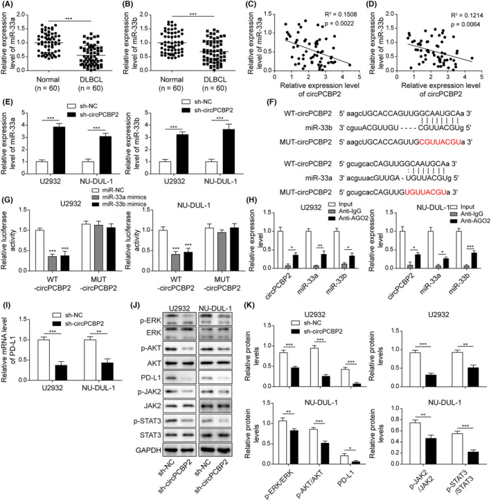
circPCBP2 directly bound with miR‐33a/b and regulated PD‐L1 signaling. (A, B) qRT‐PCR to assess miR‐33a/b levels in human DLBCL specimens. (C, D) The relationship between miR‐33a/b levels and circPCBP2 level in DLBCL tissues. (E) Relative circPCBP2 levels in sh‐NC or sh‐circPCBP2 transfected DLBCL cells. (F) Predicted binding sites between circPCBP2 and miR‐33a/b. (G) Relative luciferase activities of circPCBP2‐WT and circPCBP2‐MUT in transfected DLBCL cells. (H) Relative enrichment of circPCBP2 and miR‐33a/b following immunoprecipitation with Ago2 or IgG antibody. (I) Relative PD‐L1 mRNA levels in DLBCL cells transfected with sh‐NC or sh‐circPCBP2. (J, K) Protein levels of p‐JAK2, JAK2, p‐STAT3, STAT3, p‐ERK, ERK, p‐AKT, AKT, and PD‐L1 in transfected DLBCL cells measured by western blotting