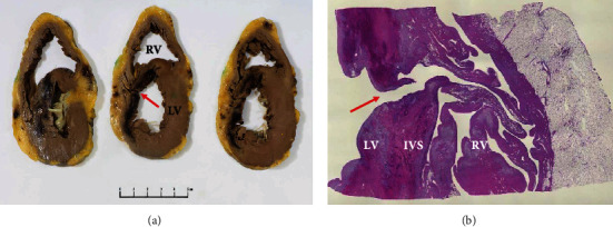
Figure 3.

Explanted heart showing. (a) Cross-section through the basal (left) to apical (right) left ventricle (LV) demonstrating haemorrhagic infarct involving the interventricular septum (IVS) and anterior LV (asterisk). A defect is evident through the infarcted region of the IVS (arrow). (b) At low power, a full thickness defect is evident through the region of infarction (arrow), allowing communication from LV to RV, via a tortuous path.
