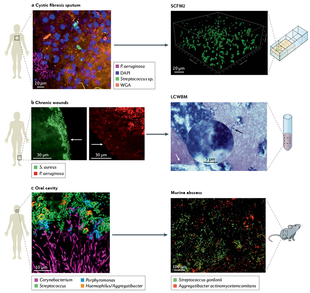
Fig. 2 |. Microbiogeography of human-associated microbial communities is the benchmark for in vitro preclinical models.

a | Spatial arrangement of Pseudomonas aeruginosa (magenta) and streptococci (green) within a cystic fibrosis sputum sample visualized using MiPACT (microbial identification after passive clarity technique) and hybridization chain reaction46. Structural context of the environment is visible using DAPI labelling of host DNA (blue) and wheat germ agglutinin (WGA) labelling of mucin (orange). Evaluation of cystic fibrosis sputum chemical composition and structure enable development of synthetic cystic fibrosis sputum medium (SCFM2), a medium that recapitulates the physical and chemical properties of cystic fibrosis sputum, where surface-detached aggregates of P. aeruginosa strain PAO1 in SCFM2 have similar spatial patterning as in cystic fibrosis sputum samples. b | P. aeruginosa (red) and Staphylococcus aureus (green) in two chronic wound biopsies visualized by peptide nucleic acid fluorescence in situ hybridization (PNA-FISH)54. Images show the spatial patterning of these two pathogens in wounds is not random. P. aeruginosa aggregates are localized deeper in wounds (50–60 μm from the surface) in comparison with S. aureus aggregates localized close to the wound surface (within 20–30 μm of the wound surface). Arrows show edge of wounds. Lubbock chronic wound biofilm model (LCWBM) provides a chemical environment similar to chronic wounds. However, spatial structure is dependent on microbial activities. Spatial arrangement of P. aeruginosa (white arrow) and S. aureus (black arrow) in this model is dependent on coagulase activity of S. aureus to turn soluble fibrinogen in plasma into insoluble fibrin, forming a fibrin network that gives gel-like properties to the media. In this model, S. aureus and P. aeruginosa can coexist in adjacent aggregates93. c | Combinatorial labelling and spectral imaging FISH (CLASI-FISH) image of microbial communities from human supragingival plaque sampled from the human oral cavity showing a spatially patterned consortia of microorganisms in complex corncob structures60. Investigations using a murine abscess model of infection demonstrated that precise spatial arrangement between Aggregatibacter actinomycetemcomitans and Streptococcus gordonii promotes virulence11. Panel a cystic fibrosis sputum adapted from REF.46, CC BY 4.0 (https://creativecommons.org/licenses/by/4.0/). Panel b chronic wound adapted with permission from REF.54, ASM. Panel b LCWBM adapted with permission from REF.93, ASM. Panel c oral cavity adapted with permission from REF.60, PNAS. Panel c murine abscess adapted with permission from REF.11, PNAS.
