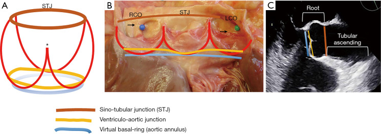
Figure 3.
The aortic root complex. (A) Schematic drawing of the aortic root: the blue line indicates the virtual basal-ring (aortic annulus); the yellow line depicts the ventriculo-aortic junction (whose non-planar nature is schematically emphasized) (33); the red lines show the crown-shaped attachments of the cusps to the wall of the aortic sinuses [note the different height of the underdeveloped commissure (pseudocommissure, asterisk) compared to that of the other 2 true commissures]; the brown line depicts the STJ. (B) All the above boundaries and structures are reported (same colors as above) in an anatomical specimen of a normal aortic root and tricuspid aortic valve. (C) Echocardiographic view of the aortic root: the levels of the aortic annulus, ventriculo-aortic junction and STJ are shown (same colors as above). It is important to recognize that it is the measurement of the virtual annulus, sinuses and STJ that have clinical and practical implications for the patient with BAV. RCO: blue pin and arrow; LCO: green pin and arrow. From Michelena et al. (1-4) with permission. STJ, sinotubular junction; BAV, bicuspid aortic valve; RCO, right coronary orifice; LCO, left coronary orifice.

