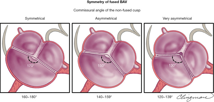
Figure 5.
Schematic of the transthoracic echocardiographic evaluation of fused BAV symmetry in the parasternal short-axis. Applicable to similar tomographic views by cardiac computed tomography and cardiovascular magnetic resonance, the figure demonstrates different commissural angles of the non-fused cusps (applicable to the 3 fused BAV phenotypes, only right-left cusp fusion is shown), which define symmetry. Left panel: symmetrical (angle 160–180°) right-left cusp fusion BAV with a raphe, where the 2 functional cusps are almost same size/shape (the non-fused cusp is a little larger), and the commissural angle of the non-fused cusp is about 170°. Middle panel: asymmetrical (angle 140–159°) right-left fusion BAV with a raphe, and the commissural angle of the non-fused cusp is about 150°. Right panel: very asymmetrical (angle 120–139°) right-left fusion BAV shows retraction of the conjoined cusp at the raphal area and the commissural angle of the non-fused cusp is about 130°. Note that retraction is more prominent as the angle decreases, which may cause aortic regurgitation. From Michelena et al. (1-4) with permission. BAV, bicuspid aortic valve.

