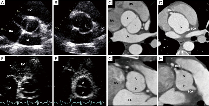
Figure 8.
Diastolic and systolic still images of the 2-sinus BAV phenotypes obtained from transthoracic echocardiogram and diastolic still images by ECG-gated cardiac CT. (A) 2-sinus laterolateral BAV in systole, with only 2 distinguishable aortic sinuses in diastole (B) and roughly equal size/shape cusps occupying 180° of the circumference, reproducible on an equivalent tomographic cut as seen with CT (C). Note the coronary arteries arising 1 from each sinus (D). (E) 2-sinus anteroposterior BAV in systole, with only 2 distinguishable aortic sinuses and roughly equal size/shape cusps occupying 180° of the circumference (F, diastolic still frame), reproducible on an equivalent tomographic cut as seen with CT (G). Note coronary arteries arising 1 from each sinus in this particular example (H). From Michelena et al. (1-4) with permission. BAV, bicuspid aortic valve; ECG, electrocardiographic; CT, computed tomography; RA, right atrium; RV, right ventricle; LA, left atrium; A, anterior cusp; P, posterior cusp; L, lateral cusp; RCA, right coronary artery; LCA, left coronary artery.

