Figures 11–18.
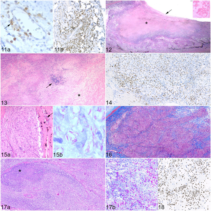
Mycobacteriosis, eye, cat. Inflammation scores: 1 = less than 1000 inflammatory cells; 2 = 1000–10,000 inflammatory cells; 3 = 10,001–50,000 inflammatory cells; 4 = 50,001–100,000 inflammatory cells; 5 = more than 100,000 inflammatory cells, calculated over the entire area of the lesion. Figure 11. Iris, Mycobacterium bovis, case 5. Inflammation score 2. (a) Lymphoplasmacytic iritis with scant T cells scattered throughout the iris stroma (black arrow). Immunohistochemistry (IHC) for CD3. (b) B cells are more abundant and outnumber T cells. IHC for Pax5. Figure 12. Ciliary body, M. bovis, case 9. Pyogranulomatous cyclitis, inflammation score 5, with extensive necrosis (asterisk) and loss of the epithelium of the pars plicata. There is a cyclitic membrane (black arrow), and Morgagnian globules in the lens (inset), indicating a cataract. Hematoxylin and eosin (HE). Figure 13. Sclera and episclera, M. bovis, case 4. Pyogranulomatous scleritis, inflammation score 5, with degeneration of collagen (asterisk) and a cluster of lymphocytes (black arrow). HE. Figure 14. Sclera, Mycobacterium lepraemurium, case 22. Pyogranulomatous scleritis, inflammation score 4, with “atypical” granulomas showing immunolabeled monocytes and granulocytes, mostly within the granuloma; macrophages and epithelioid macrophages are negative. IHC for calprotectin. Figure 15. Corneal limbus, M. lepraemurium, case 21. Inflammation score 3. (a) Pyogranulomatous perilimbal inflammation expanding the stroma, with the formation of non-necrotic pyogranulomas dominated by epithelioid macrophages. Normal features of the corneal limbus such as blood vessels (black arrow) and melanocytes are present. HE. (b) Positive staining for acid-fast bacilli indicative of mycobacteria within pyogranulomas. Bacterial index grade 5. Ziehl-Neelsen (ZN). Figure 16. Conjunctiva, Mycobacterium microti, case 3. Pyogranulomatous conjunctivitis of the third eyelid, inflammation score 5. Collagen fibers are present, subdividing the inflammatory infiltrate. The fibrous capsules are thin and incomplete. Masson’s trichrome. Figure 17. Optic nerve, M. bovis, case 8. Inflammation score 5. (a) Pyogranulomatous optic neuritis with necrosis (asterisk). HE. (b) Abundant acid-fast bacilli (pink) present within the optic nerve. The overall bacterial index grade was 5. ZN. Figure 18. Vitreous, M. bovis, case 17. Neutrophils are numerous within the vitreous. IHC for calprotectin.

