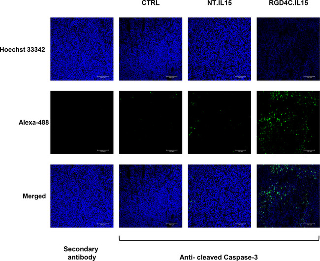
Figure EV5. Immunostaining of tumour sections using an anti‐cleaved caspase‐3 antibody.

Tumour sections from tumour‐bearing mice (n = 6) following single TPA.IL15 IgK dose treatment, 5 × 10¹⁰ TU/mouse, of targeted (RGD4C.IL15) or non‐targeted (NT.IL15). Untreated mice were used as controls (CTRL). Tumour sections incubated with the secondary antibody alone were also included as negative controls. Hoechst 33342 was used to stain the cell nuclei. Scale bar, 100 μm. Source data are available online for this figure.
