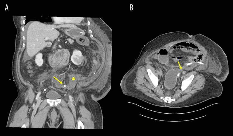
Figure 2.
CT scans of rectal stump dehiscence with free fluid. A shows free fluid accumulation in the pelvis secondary to the rectal stump dehiscence in the coronal view. The placement of the Blake drain is indicated by the yellow arrow, and the collection of free fluid is indicated by the yellow star. B shows the rectal stump dehiscence in the transverse view. The rectal stump dehiscence is indicated by the yellow arrow.

