
FIGURE 7.
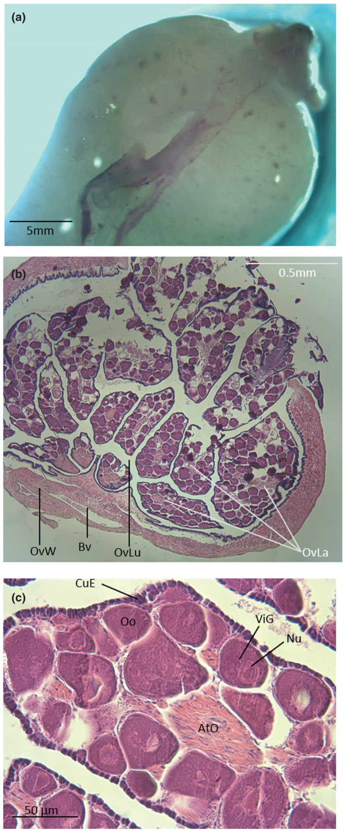
Dorsal view (a) and cross sections (b and c) of an ovary in Parophidion vassali. Ato, Atretic oocyte; Bv, Blood vessel; CuE, Cubic epithelium; nu, Nucleolis; Oo, Oocyte; OvLa, Ovarian lamella; OvLu, Ovisac lumen; OvW, Ovisac wall; ViG, Vitelline granule.