
FIGURE 10.
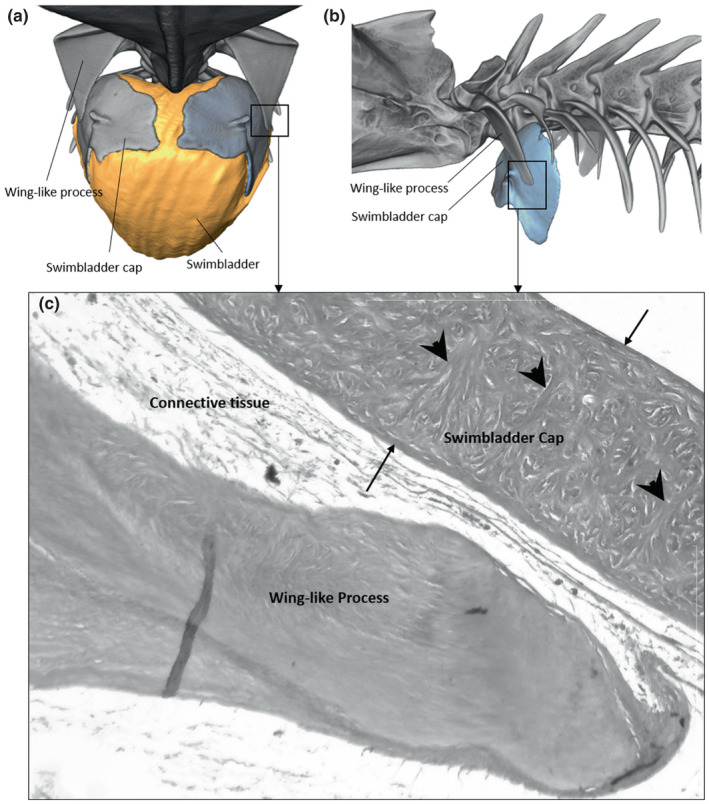
Frontal (a) and left lateral views (b) of the swimbladder cap in Parophidion vassali. Histological cross section (c) showing the swimbladder cap and distal tip of the wing‐like process. The swimbladder cap is composed of two external wall (arrow) of fibres that are connected by transversal fibres (arrowhead). Between the fibres are found mineralised items (white spaces).