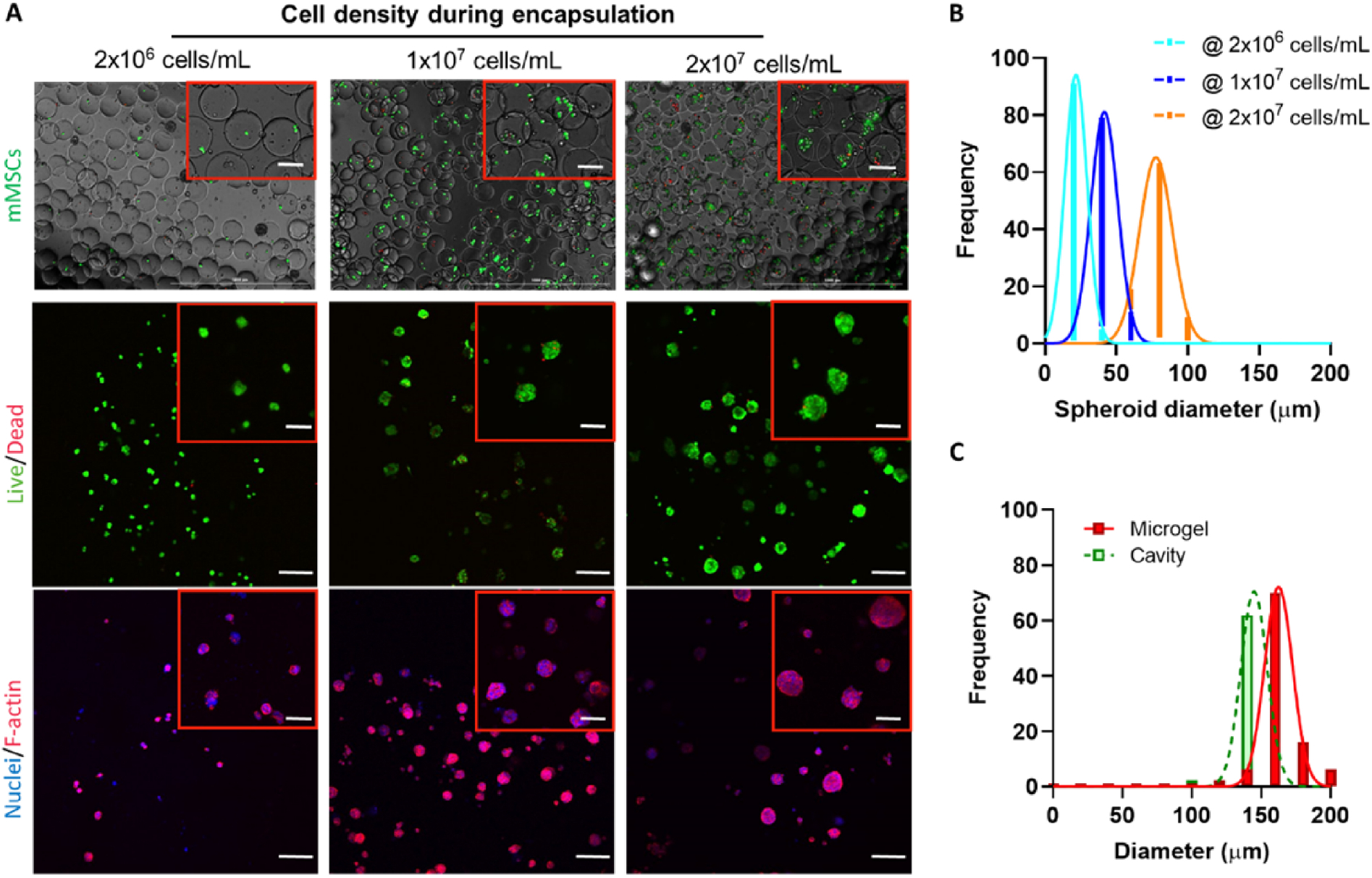
Figure 4. mMSC solid spheroid formation within macroporous hydrogels.

(A) Live/Dead staining of mMSCs immediately after microencapsulation into PEGNB-Dopa microgels. mMSCs with a density of 2×10⁶ cells/mL, 1×10⁷ cells/mL, and 2×10⁷ cells/mL in OptiPrep were used for encapsulation. (B) Live/Dead and cytoskeleton fluorescence staining of mMSC spheroids after 7 days of culture. Scale bar = 100 μm. (C) Quantification of the sizes of microgels, cavities, and mMSC spheroids formed with various cell loading concentrations after 7 days of culture.
