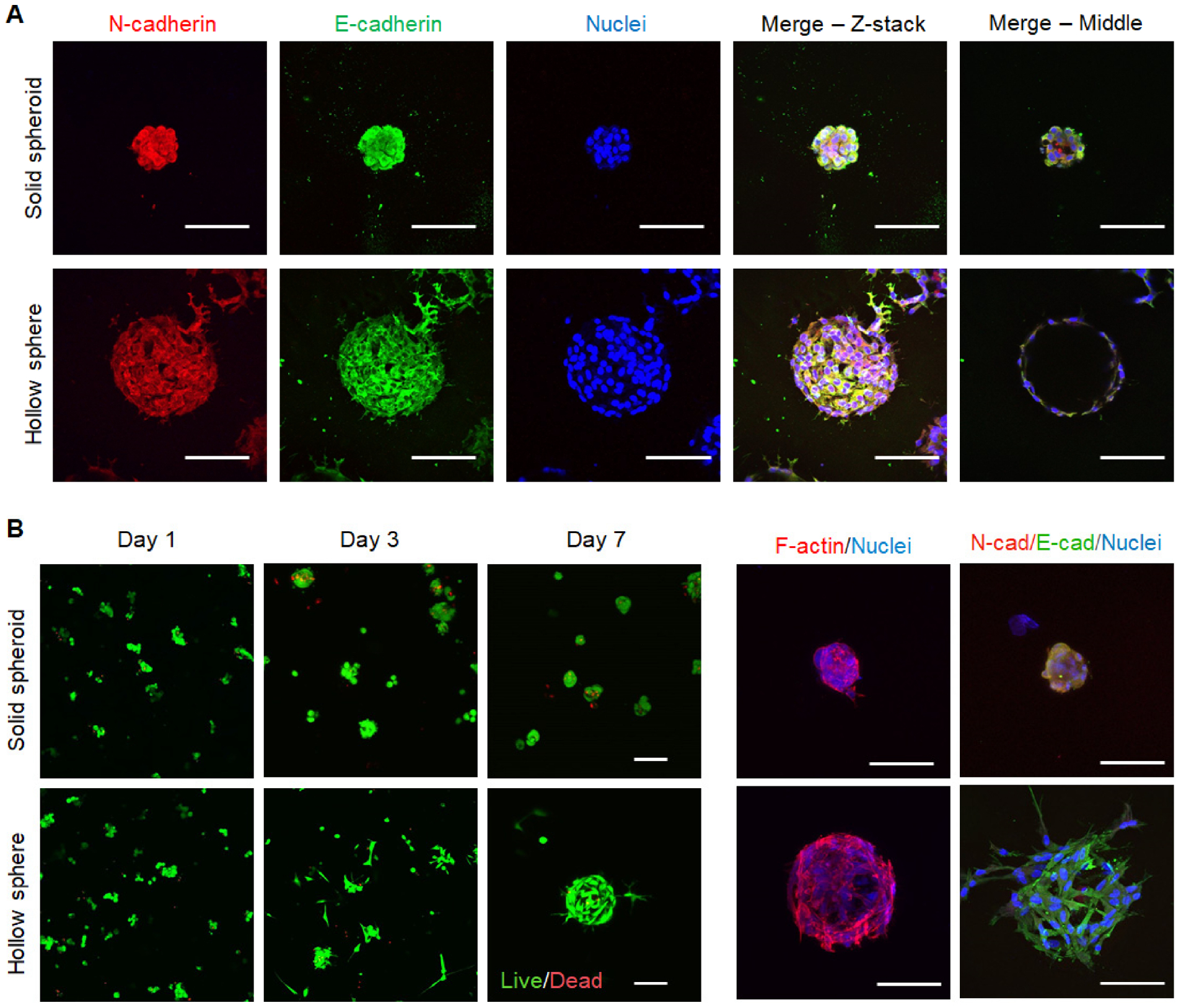
Figure 5. Fluorescence imaging of solid spheroids and hollow spheres formed with MSCs.

(A) Immunofluorescence staining of E/N-cadherin and nuclei in mMSC solid spheroids or hollow spheres on day 7. (B) Live/Dead staining and immunofluorescence staining of cytoskeleton, E/N-cadherin and nuclei in hMSC solid spheroids or hollow spheres. Scale bar = 100 μm.
