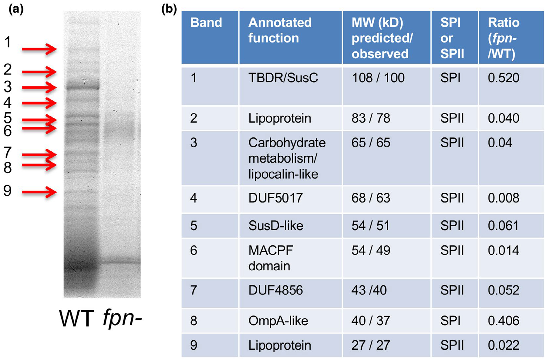
FIGURE 3.

The level of many secretome components is strongly reduced in an fpn− strain. (a) Proteins present in the culture medium of wild-type (WT) ETBF and isogenic fpn− strains were resolved by SDS-PAGE and visualized using Colloidal Blue. (b) Nine proteins that were enriched in the culture medium of wild-type cells (red arrows) were excised from the gel and identified by mass spectrometry. The predicted size of each protein (lacking the signal peptide) and the observed size (based on mobility on SDS-PAGE) is indicated. The presence of a signal peptidase I (SP I) or signal peptidase II (SP II) cleavage site as predicted by LipoP is shown. The ratio of each protein in the secretome of the two strains is based on an analysis of the culture media by quantitative mass spectrometry
