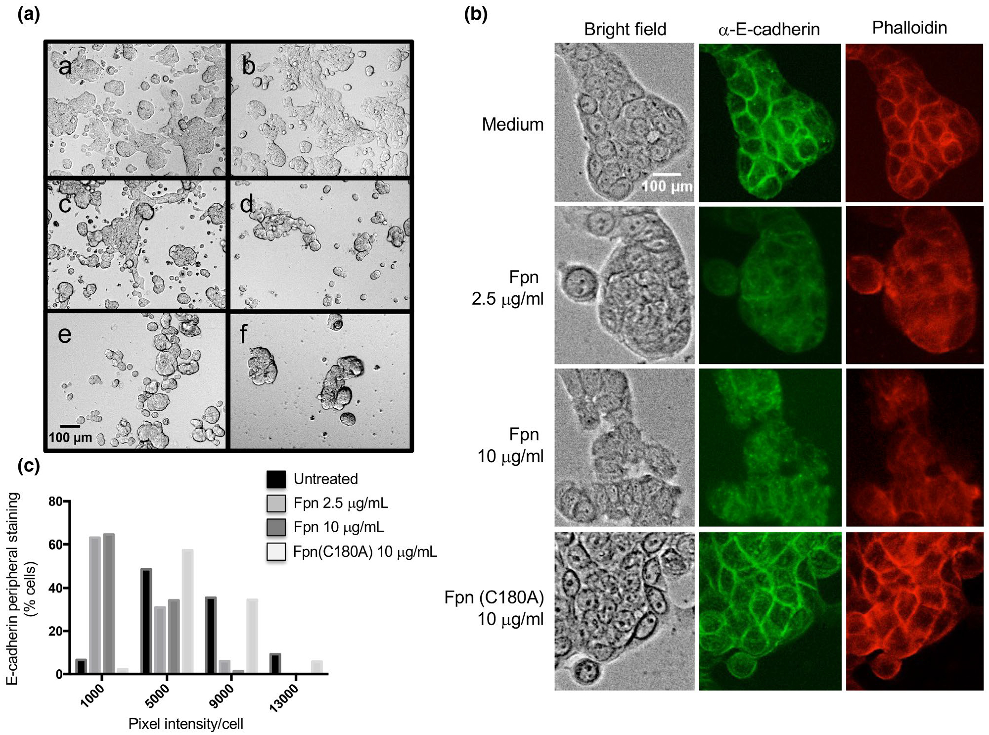
FIGURE 7.

Incubation of HT29 cells with Fpn leads to a loss of adhesion and cell-cell junctions. (A) Live cell microscopy (bright field, 10X magnification) of HT29 cells incubated with culture medium alone (a), 10 μg/ml Fpn (C180A) (b), 2.5 μg/ml Fpn (c), 10 μg/ml Fpn (d), 2.5 μg/ml C. histolyticum Clo protease (e), or 10 μg/ml Clo (f). (B) HT29 cells were incubated with culture medium alone, Fpn (2.5 or 10 μg/ml) or Fpn (C180A) (10 μg/ml). Cells were fixed, stained with either FITC-conjugated anti-E-cadherin or rhodamine-conjugated phalloidin, and visualized at 20X magnification. (C) The intensity of peripheral E-cadherin staining in individual cells was determined, and the results were binned based on the percent of cells within the population that showed different levels of staining (arbitrary units)
