Fig. 1.
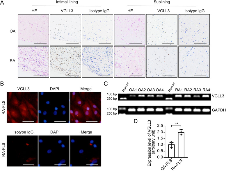
Expression of VGLL3 in the synovial tissues of rheumatoid arthritis (RA) and osteoarthritis (OA) patients. a Representative images of the expression of VGLL3 examined by immunohistochemistry staining using anti-VGLL3 antibodies and rabbit IgG (isotype) in RA and OA synovial tissues. Hematoxylin and eosin (HE) staining was also performed to evaluate the pathological changes. N = 5. Scale bar, 500 μm. b Representative images of the expression of VGLL3 in the fibroblast-like synoviocytes (FLS) in RA examined by immunofluorescence staining. Nuclei were stained with DAPI. N = 3. Scale bar, 50 μm. c The mRNA expression of VGLL3 in RA-FLS and OA-FLS was detected by reverse transcription PCR (RT-PCR). GAPDH was used as the housekeeping gene. N = 4. d The mRNA expression levels of VGLL3 in RA-FLS and OA-FLS were detected by real-time quantitative PCR (qPCR). N = 3, ** p < 0.01. Data were shown as mean ± standard deviation (SD)

