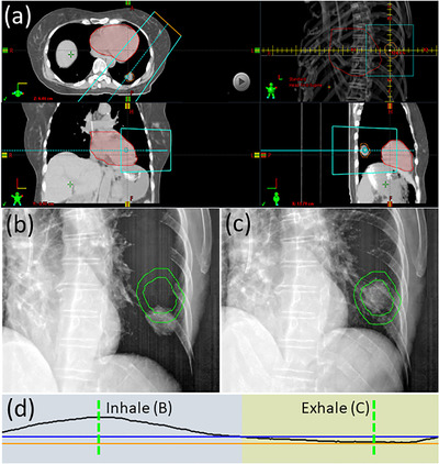
FIGURE 3.

Example of a lung tumor that can be visualized in fluoroscopy with an oblique projection angle. (a) Axial, coronal, and sagittal average CT images, demonstrating that the tumor is positioned posterior to the heart, which will obscure visualization in anterior–posterior projection fluoroscopy. (a) also depicts the beam's eye view of an oblique angled fluoroscopic imaging field, where separation can be observed between the heart (red) and ITV (orange) structures. (b) The ITV and PTV contours (green) and the tumor position on both (b) inhale and (c) exhale oblique fluoroscopy. The corresponding amplitudes of the breathing trace for these images are shown by the dotted green lines in (d). Using fluoroscopy, the gating window (orange and blue lines) is set so that beam‐on (indicated by yellow colorwash) will only occur while the tumor is inside the ITV. This patient is an example of a Gate4060 treatment
