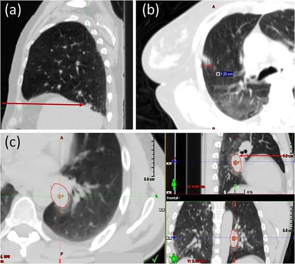
FIGURE 5.

Example cases where thoracic tumors are not visible in fluoroscopic imaging. (a) Sagittal CT image showing tumor located posterior to the diaphragm and inferior to the apex of the diaphragm, such that diaphragm will obscure tumor visualization in an anterior–posterior projection. (b) Axial CT image showing a diffuse tumor, or ground glass object, with signal that is too faint to be observed in fluoroscopy. (c) Axial, sagittal, and coronal CT images of a tumor that is located posterior to mediastinal soft tissue, such as the heart
