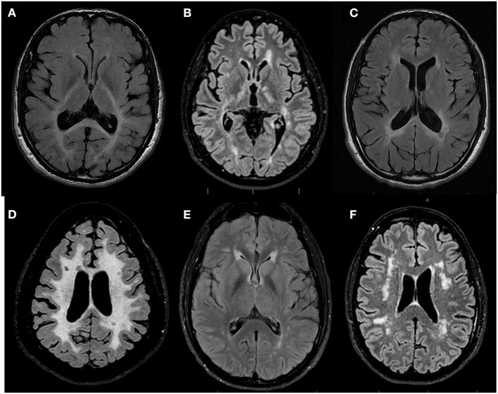
Figure 3.
Representative Brain MRI of patients referred to the WM Rounds according to their pattern of involvement. Representative axial FLAIR T2-weighted brain MR images of patients referred to the WMR according to their pattern of involvement. (A) 44-year-old man finally diagnosed with Peroxisomal Biogenesis Disorder showing confluent symmetric white matter abnormalities predominant in the posterior regions and affecting the splenium of the corpus callosum and the posterior limb of the internal capsules; (B) 29-year-old man diagnosed with X-linked Charcot-Marie-Tooth Disease and MS showing multifocal white matter lesions compatible with MS plaques; (C) 43-year-old man with suspected genetic MS mimicker showing bilateral periventricular white matter changes with involvement of the splenium of the corpus callosum and focal T2 hypointense lesions adjacent to the periventricular white matter; (D) 44-year-old man (no final diagnosis) with confluent symmetric white matter involvement in which focal T2-hypointense lesions suggestive of MS plaques were identified; (E) 38-year-old man with CADASIL (patient vignette no. 1) showing multifocal white matter abnormalities and anterior symmetric periventricular involvement; (F) 58-year-old woman (patient vignette no. 2) showing multifocal lobar white matter abnormalities.

