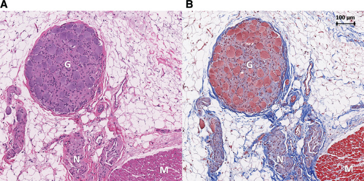
Figure 5:
Treatment site histology. Histology of an atrial section at the treated oblique sinus ganglionated plexus region in animal #4 prepared with hematoxylin and eosin (A) as well as Masson’s trichome (B) stains. Nerve fibers (N) and a ganglion (G) are shown within epicardial fat adjacent to atrial myocardium (M). Tissue architecture is preserved without cellular disarray or nuclear disintegration.

