. 2022 Mar 15;82(6):1013–1024. doi: 10.1158/0008-5472.CAN-21-1707

Figure 3.

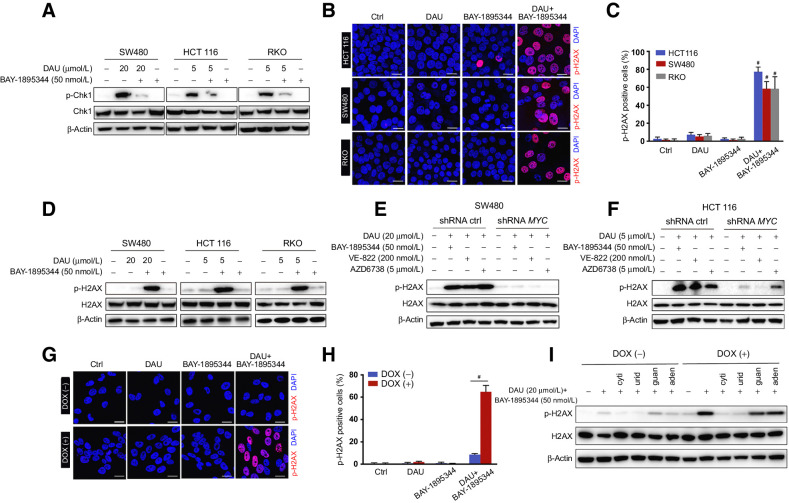
Figure 3. DAU combined with ATR inhibitors induces DNA damage in a MYC-dependent manner. A, Immunoblots of the indicated proteins in SW480, HCT116, and RKO cells treated for 24 hours with DAU alone or together with BAY-1895344. B, Phosphorylation of H2AX in HCT116, SW480, and RKO cells treated with DAU and BAY-1895344 alone or together were revealed by immunostaining. Phospho-H2AX (14) and DAPI (blue). Quantified data are shown in C. #, P < 0.0001; significantly different from vehicle-treated group. D, Immunoblots of the indicated proteins in SW480, HCT116, and RKO cells treated with DAU and BAY-1895344 alone or together for 24 hours. E and F, Immunoblots of the indicated proteins in control and MYC knockdown SW480 (E) and HCT116 (F) cells treated with DAU alone or together with BAY-1895344, VE-822, or AZD6738 for 24 hours. G, Phosphorylation of H2AX in ARPE-19-MYC (DOX+) and ARPE-19-MYC (DOX−) cells treated with 20 μmol/L DAU and 50 nmol/L BAY-1895344 alone or together for 16 hours. Quantified data are shown in H. #, P < 0.0001. I, Immunoblots of the indicated proteins in ARPE-19-MYC (DOX+) and ARPE-19-MYC (DOX−) cells treated with DAU and BAY-1895344 with or without 50 μmol/L cytidine (cyti), uridine (urid), guanosine (guan), or adenosine (aden). Graphic data are means ± SEM. For Western blotting, one of three to five similar experiments is shown. Scale bar, 20 μm. Ctrl, control; DOX, doxycycline.

DAU combined with ATR inhibitors induces DNA damage in a MYC-dependent manner. A, Immunoblots of the indicated proteins in SW480, HCT116, and RKO cells treated for 24 hours with DAU alone or together with BAY-1895344. B, Phosphorylation of H2AX in HCT116, SW480, and RKO cells treated with DAU and BAY-1895344 alone or together were revealed by immunostaining. Phospho-H2AX (14) and DAPI (blue). Quantified data are shown in C. #, P < 0.0001; significantly different from vehicle-treated group. D, Immunoblots of the indicated proteins in SW480, HCT116, and RKO cells treated with DAU and BAY-1895344 alone or together for 24 hours. E and F, Immunoblots of the indicated proteins in control and MYC knockdown SW480 (E) and HCT116 (F) cells treated with DAU alone or together with BAY-1895344, VE-822, or AZD6738 for 24 hours. G, Phosphorylation of H2AX in ARPE-19-MYC (DOX+) and ARPE-19-MYC (DOX) cells treated with 20 μmol/L DAU and 50 nmol/L BAY-1895344 alone or together for 16 hours. Quantified data are shown in H. #, P < 0.0001. I, Immunoblots of the indicated proteins in ARPE-19-MYC (DOX+) and ARPE-19-MYC (DOX) cells treated with DAU and BAY-1895344 with or without 50 μmol/L cytidine (cyti), uridine (urid), guanosine (guan), or adenosine (aden). Graphic data are means ± SEM. For Western blotting, one of three to five similar experiments is shown. Scale bar, 20 μm. Ctrl, control; DOX, doxycycline.