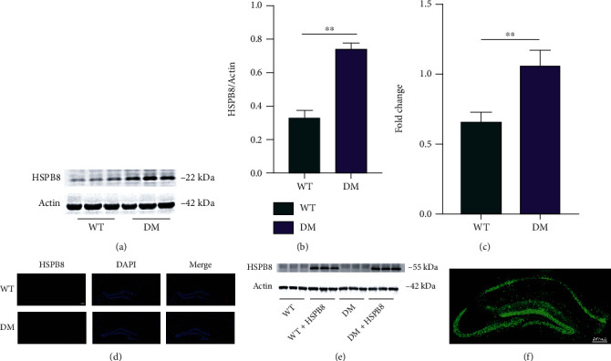
Figure 1.

Upregulation of HSPB8 in the hippocampus of DM mice. (a, b) Representative western blot images and quantitative analyses of HSPB8 in the hippocampus of WT and DM mice (n = 5 mice per group). ∗∗, p < 0.01. (c) mRNA levels of HSPB8 in the hippocampus of WT and DM mice were detected by PCR. ∗∗, p < 0.01. (d) Representative immunofluorescence staining images of HSPB8 (red) in the hippocampus of WT and DM mice. Nuclei were counterstained with DAPI (blue). Scale bar = 200 μm. (e) Upregulation of HSPB8 was confirmed by western blotting and (f) immunofluorescent staining when injected virus in the hippocampus (n = 5 mice per group).
