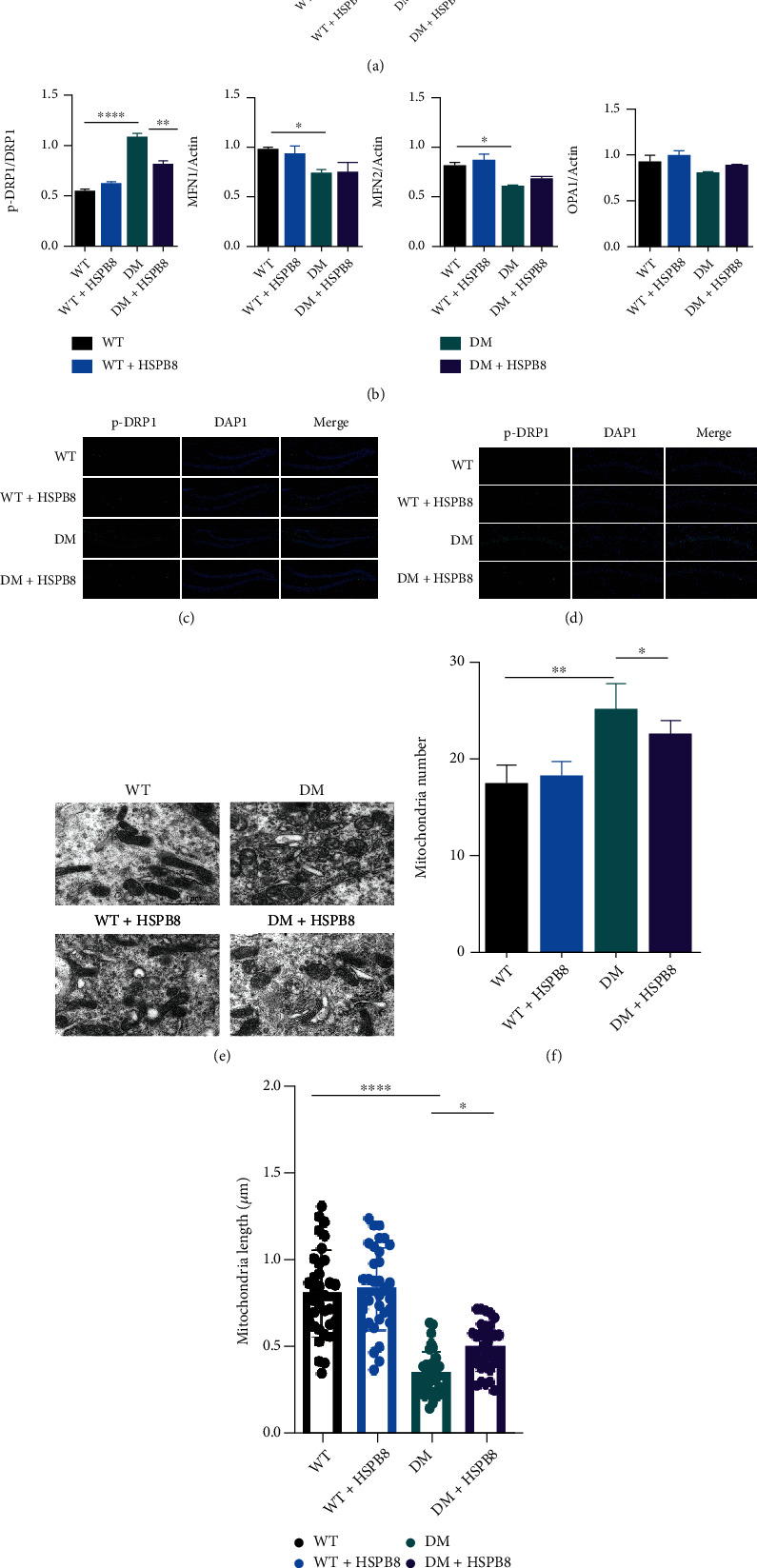
Figure 4.

HSPB8 overexpression in the hippocampus alleviates mitochondrial fission. (a) Expressions of p-Drp1, Drp1, MFN1, MFN2, and OPA1 in the hippocampus were detected by Western blot. β-Actin was used as loading control. (n = 5 each group) and (b) quantitative analysis. ∗, p < 0.05, ∗∗, p < 0.01, ∗∗∗∗, p < 0.0001. (c, d) Representative images of immunofluorescence staining against p-Drp1 (green) in the (dentate gyrus) DG and CA1. Nuclei were counterstained with DAPI (blue) (n = 3 mice per group). Scale bar = 100 μm. (e) Representative electron microscopy images showing mitochondrial morphology in hippocampal neurons (n = 3 mice per group). Scale bar = 1 μm. (f, g) Measurements of mitochondrial numbers and mitochondrial length. One-way ANOVA test followed by Tukey's post hoc test. Data are shown as the mean ± SEM. ∗p < 0.05, ∗∗, p < 0.01.
