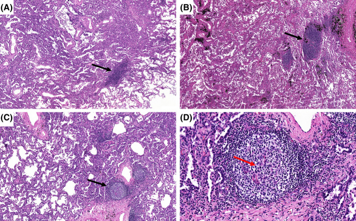
FIGURE 2.

Histological appearance of intra‐tumoral TLSs. (A) Grade I TLSs: Aggregates are vague, ill‐defined clusters of lymphocytes (black arrow: HE×100). (B) Grade II TLSs: Primary follicles consist of dense, round, or oval shaped clusters of lymphocytes (black arrow: HE ×100). (C) Grade III TLSs: Secondary follicle are centered by a germinal center (black arrow: HE ×100). (D) Grade III TLSs: At high magnification, microscopic examination of a secondary follicle shows a pale area (germinal center‐red arrow), with a dense outer rim of lymphocytes (mantle zone) (HE ×400). HE, hematoxylin and eosin stain; TLSs, tertiary lymphoid structures. (This figure appears in color on the web).
