
FIGURE 6.
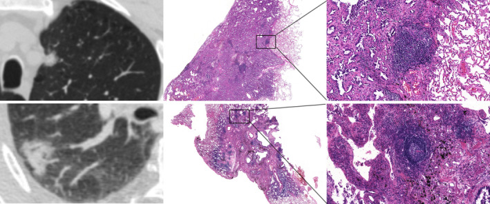
CT images and corresponding HE histologic findings in peripheral LAC characterized by SN. Top row: images in a 64‐year‐old man with LAC. CT shows SN and the corresponding pathological manifestation is grade II semi‐mature TLSs in tumor tissues. Bottom row: images in a 61‐year‐old woman with LAC. CT shows SN and the corresponding pathological manifestation is grade III mature TLSs in tumor tissues. CT, computed tomography; HE, hematoxylin and eosin stain; LAC, lung adenocarcinoma; SN, solid nodule; TLSs, tertiary lymphoid structures.