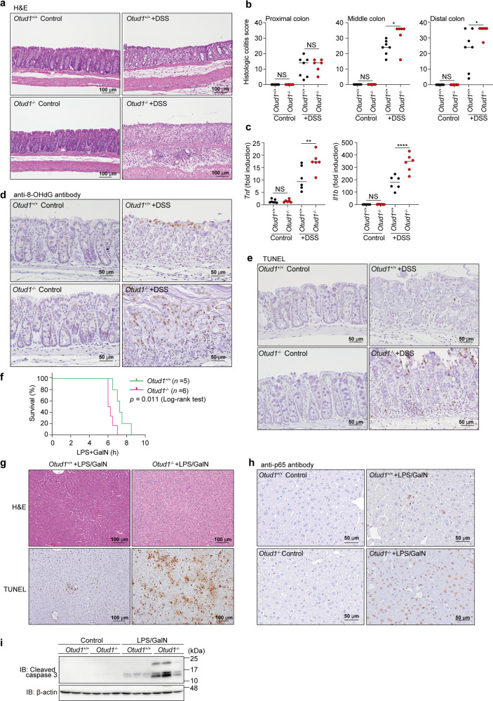
Fig. 6. Enhanced inflammation, oxidative damage, and cell death in mouse inflammatory disease models.
a, b Colon damage in DSS-administered Otud1−/−-mice. Otud1+/+- and Otud1−/−-mice were treated with 2.5% DSS for 7 days and then sacrificed. Representative images of H&E staining of distal colon (a) and histologic scores of colitis b (n = 6–7) are indicated. Bars; 100 µm. c Increased expression of NF-κB target genes in DSS-treated Otud1−/−-mice. qPCR analysis of NF-κB targets in the distal colon from DSS-treated mice (n = 6). Data are shown as scatter plots and evaluated by Mann–Whitney test. *P < 0.05, **P < 0.01, ****P < 0.0001, NS: not significant. d, e Increased oxidative DNA damage and cell death in DSS-administered Otud1−/−-mice. Specimens as in a were stained for 8-OHdG (d) or TUNEL (e). Bars: 50 µm. f Otud1−/−-mice were labile for the LPS/GalN-induced acute hepatitis model. After injections of LPS (10 μg/kg) and GalN (800 mg/kg), the % survivals of Otud1+/+-(n = 5) and Otud1−/−-(n = 6) mice were determined by the Kaplan–Meier method with the Log-rank test. g Enhanced liver damage in Otud1−/−-mice. H&E and TUNEL staining of livers from Otud1+/+- and Otud1−/−-mice after 5 h administration of LPS/GalN. Bars: 100 µm. h Increased intranuclear localization of p65 in hepatocytes from LPS/GalN-treated Otud1−/−-mice. The specimens in g were stained with an anti-p65 antibody. Bars: 50 µm. i Increased caspase 3 activations in hepatocytes from Otud1−/−-mice. After a 5 h treatment with LPS/GalN, livers were excised, lysed, and immunoblotted with the indicated antibodies.

