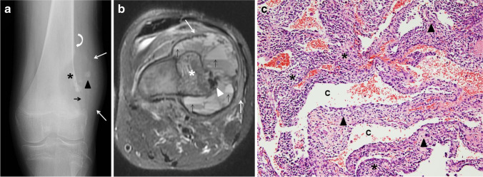
Fig. 10.
Telangiectatic osteosarcoma in a 17-year-old boy. a An initial anteroposterior radiograph of the knee shows a distal femoral metadiaphyseal, geographical, eccentric, lytic lesion (asterisk) with no sclerotic border. There is focal destruction of the distal femoral cortex (black arrow), focal areas of mineralization (arrowhead) and an associated soft-tissue mass (white arrows). Codman’s triangle is present proximally (curved arrow). b An axial fat-suppressed T2-weighted magnetic resonance image of the femoral lesion shows the eccentric, osseous mass (asterisk) with multiple fluid-fluid levels located in the periphery (black arrows), hypointense areas of mineralization (arrowhead) and adjacent soft tissue edema (white arrows). c Hematoxylin and eosin staining, magnification 100X: solid areas and cystic spaces (C) filled with blood, fibrous cellular septa (arrowheads), malignant stromal cells (asterisks)

