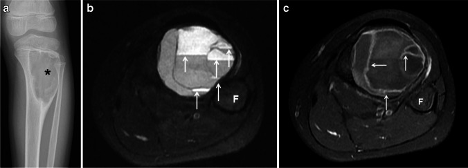
Fig. 3.
Type III aneurysmal bone cyst of the tibia in a 10-year-old girl. a An anteroposterior radiograph of the tibia shows an eccentric, well-defined, juxtaphyseal, lytic and expansile lesion (asterisk) involving the proximal tibial metadiaphysis. b An axial fat-suppressed T2-weighted magnetic resonance (MR) image of the leg shows fluid-fluid levels throughout the lesion (arrows). c A fat-suppressed, contrast-enhanced, T1-weighted MR image of the lesions shows thin, enhancing septations (arrows). F fibula

