Fig. 5.
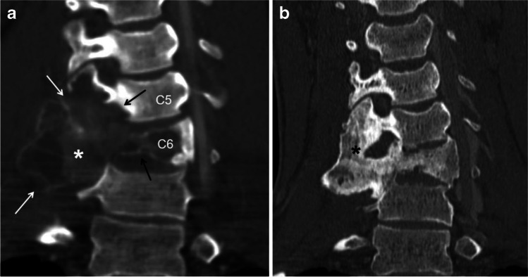
Aneurysmal bone cyst (ABC) of the cervical spine involving two consecutive vertebrae in a 16-year-old boy before and after percutaneous sclerotherapy with doxycycline. a A coronal reconstruction of a contrast-enhanced computed tomography (CT) scan image of the cervical spine using bone window shows a large, expansile lytic lesion (asterisk) involving the transverse processes (white arrows), articular pillars and body of C5 and C6 (black arrows). b A coronal reconstruction of a non-enhanced CT scan image of the cervical spine using bone window 6 years later after sclerotherapy shows complete interval healing of the lesion (asterisk) with diffuse sclerosis, remodeling of the bone and localized fusion at the site of the treated ABC

