Fig. 6.
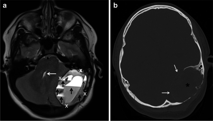
Aneurysmal bone cyst (ABC) of the skull and petrous bone in a 10-year-old boy. a An axial T2-weighted magnetic resonance image of the head shows a large, elliptical, expansile mass (calipers) in the left side of the posterior fossa causing mass effect upon the left cerebellar hemisphere and partial effacement of the fourth ventricle (white arrow). The mass is multiseptated and contains fluid-fluid levels of different signal intensity distributed throughout the lesion (black arrows). b An axial computed tomography scan image using bone windows shows to better advantage the petrous bone and occipital bone involvement by the ABC (asterisk) as well as a thin peripheral calcified shell (white arrows)

