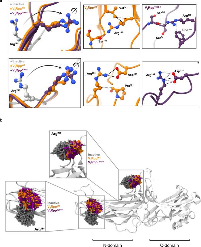
Fig. 3. Structural insights into binding of phospho-peptides to βarr1.
a Structural snapshots comparing the relative orientation and local interaction networks of trypsin cleavage sites Arg188 and Arg285 in the crystal structures of βarr1 in basal (PDB: 1G4M, grey), V2RppWT-bound (PDB: 4JQI, orange) and V2RppT360-1-bound (PDB: 7DFA, violet) conformations. The dotted lines represent hydrogen bonds and polar interactions. b Molecular dynamics simulations based on the crystal structures confirm an overall similar conformational space sampled by Arg188 and Arg285, the two trypsin proteolysis sites which are protected by ScFv30.

