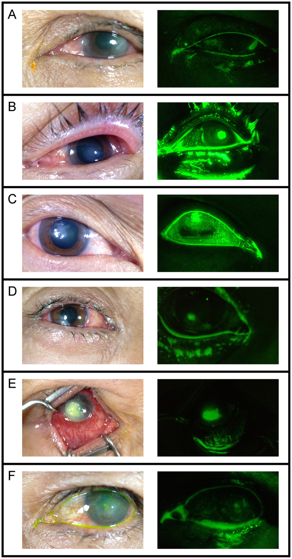
Figure 2: Corneal Photographs of Participants Diagnosed with an Epithelial Defect by the On-site Ophthalmologist.

Photographs of the 6 subjects found to have epithelial defects (white arrowheads) taken with the white light smartphone attachment (left) and the fluorescein smartphone attachment (right). When viewed as a pair, all three graders correctly identified the epithelial defects in panels A through E and one grader correctly identified the epithelial defect in panel F.
