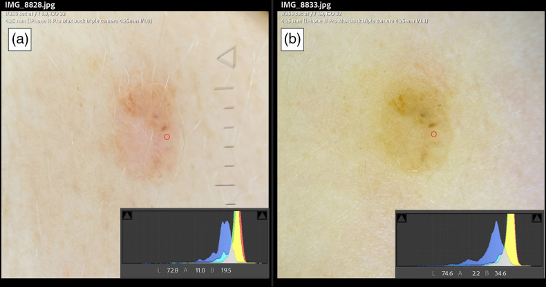
Fig. 7.
A real-world scenario of differences in dermoscopes LED color tint showing the same pigmented lesion and standard exposure settings. Panel A is acquired using a dermoscope with a slightly pink shift using an iPhone 11 with its corresponding histogram and a point localized on the lesion with a red circle and the corresponding L*a*b* values. Panel B using a different dermoscope with more of a yellow hue, its corresponding histogram, and a red circle in the same localization showing L*a*b* values of this point.

