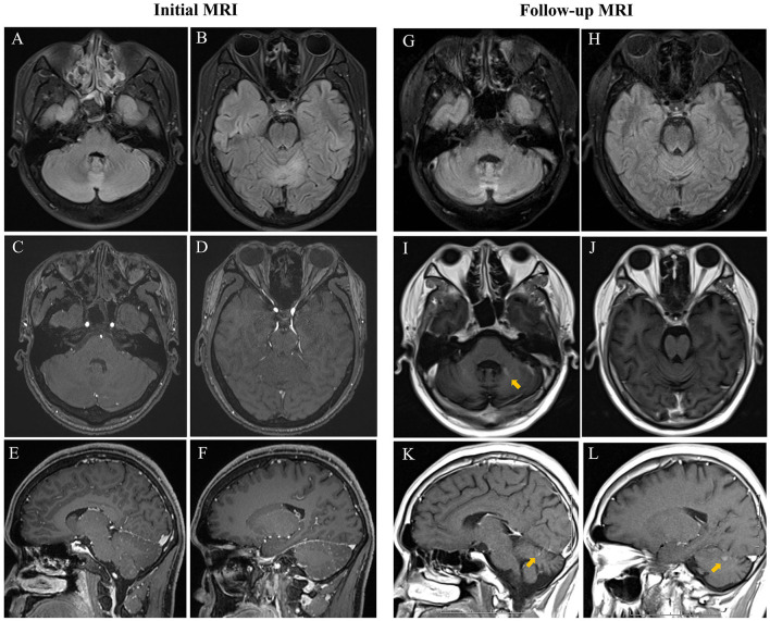
Figure 1.
Initial cerebral MRI showed increased signal in the vermis and both bilateral cerebellar hemispheres on FLAIR (A,B) without enhancement on contrast-enhanced T1-weighted sequence (C–F). Repeated MRI showed an increase of FLAIR hyperintensity of cerebellar lesions (G) and progression in size of the vermis lesions (H) with slightly enhanced T1-weighted signals on both axial and sagittal view and obvious cerebellar atrophy (I–L).

