
Figure 2.
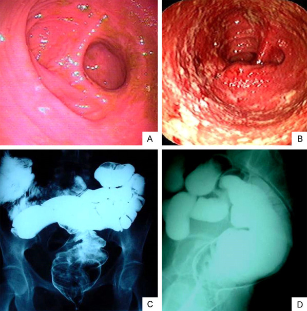
Endoscopic and radiological aspects of the ileal pouch. Endoscopic image of patients showing in (A) Normal mucosal appearance and in (B) Abnormal mucosa suggesting pouchitis. The endoscopic elements of the ileal pouchitis are granularity, loss of vascular pattern, edema, friability, mucosal hemorrhage, and superficial ulcers (Endoscopy Unit, Gastrocenter, Unicamp). Radiological aspects of the ileal pouch in the pouchogram, in (C) Frontal view (posteroanterior) and (D) Lateral view evidencing no abnormalities performed before ileostomy closure (Radiology Unit, Unicamp).