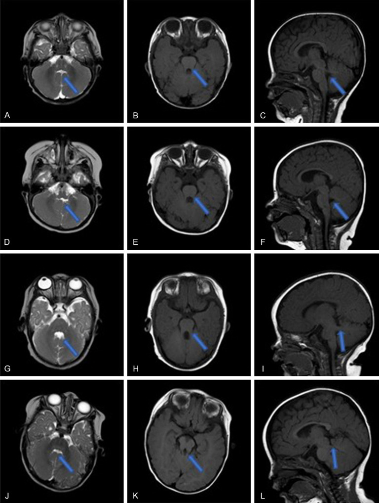
Figure 1.

MRI results of the 4 patients. A-C. Patient 1. A. T2WI coronal plane: line-like cerebrospinal fluid between the bilateral cerebellar hemispheres with long T2 signal “midline fissure sign”, the upper part of the fourth ventricle “bat wing”. B. T1WI coronal plane: the midbrain and the enlarged upper cerebellar peduncle form a “grinding sign”. C. T1WI sagittal plane: absence of inferior cerebellar vermis. D-F. Patient 2. D. T2WI coronal plane: line-like cerebrospinal fluid between the bilateral cerebellar hemispheres with long T2 signal “midline fissure sign”, the upper part of the fourth ventricle “bat wing”. E. T1WI coronal plane: the midbrain and the enlarged upper cerebellar peduncle form a “grinding sign”. F. T1WI sagittal plane: absence of inferior cerebellar vermis. G-I. Patient 3. G. T2WI coronal plane: the upper part of the fourth ventricle is “bat wing-shaped”, and the middle part expands into a triangle. H. T1WI coronal plane: the midbrain and the enlarged upper cerebellar peduncle form a “grinding sign”. I. T1WI sagittal plane: the enlarged upper cerebellar peduncle is almost perpendicular to the midbrain. J-L. Patient 4. J. T2WI coronal plane: line-like cerebrospinal fluid between the bilateral cerebellar hemispheres with long T2 signal “midline fissure sign”, the upper part of the fourth ventricle “bat wing”. K. T1WI coronal plane: the midbrain and the enlarged upper cerebellar peduncle form a “grinding sign”. L. T1WI sagittal plane: the enlarged upper cerebellar peduncle is almost perpendicular to the midbrain.
