. 2022 Jul 15;4(4):e210076. doi: 10.1148/rycan.210076

Figure 1:

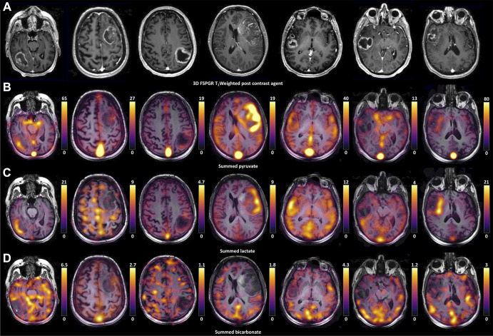
Hyperpolarized 13C MR images from all seven patients. (A) Grayscale axial contrast-enhanced 1H three-dimensional (3D) T1-weighted fast spoiled gradient-echo (FSPGR) images through the center of the lesion for each patient and the corresponding unenhanced images overlaid with the (B) pyruvate, (C) lactate, and (D) bicarbonate color maps summed over the time course.

Hyperpolarized 13C MR images from all seven patients. (A) Grayscale axial contrast-enhanced 1H three-dimensional (3D) T1-weighted fast spoiled gradient-echo (FSPGR) images through the center of the lesion for each patient and the corresponding unenhanced images overlaid with the (B) pyruvate, (C) lactate, and (D) bicarbonate color maps summed over the time course.