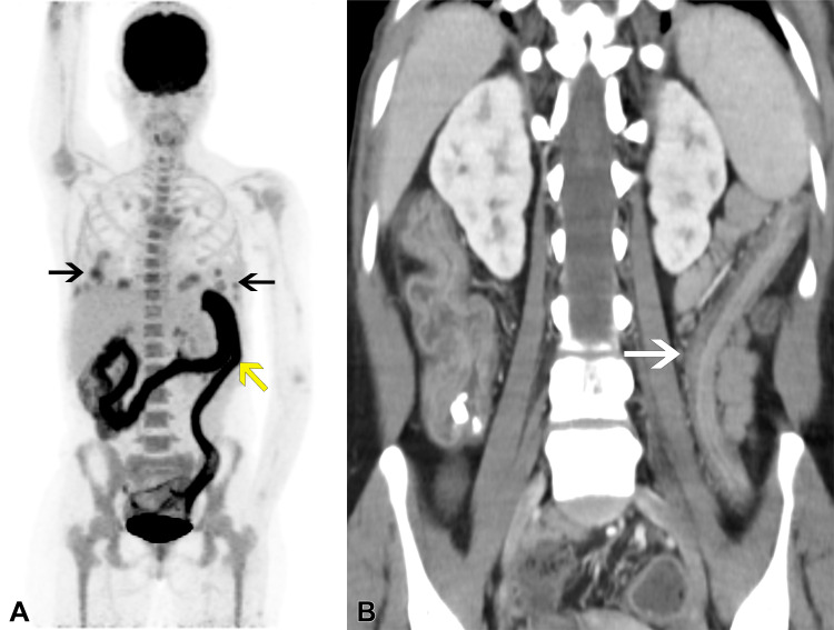
Figure 2:
Follow-up imaging in the same patient after tacrolimus withdrawal and initiation of rituximab induction therapy. Despite initial improvement, the patient presented with fever, abdominal pain, and bloody diarrhea after 2 months, before starting chemotherapy. (A) PET/CT image reveals improved uptake levels of the pulmonary masses (black arrows), but intense, diffuse, pancolonic fluorodeoxyglucose uptake was detected, compatible with active colitis (yellow arrow). (B) Corresponding coronal CT image reveals characteristic “lead-pipe” colon (white arrow), indicating clinical suspicion of ulcerative colitis exacerbation secondary to rituximab. Laboratory test results ruled out infection. Low-dose corticosteroid treatment was started with immediate clinical improvement.

