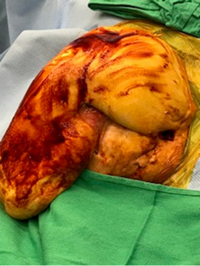
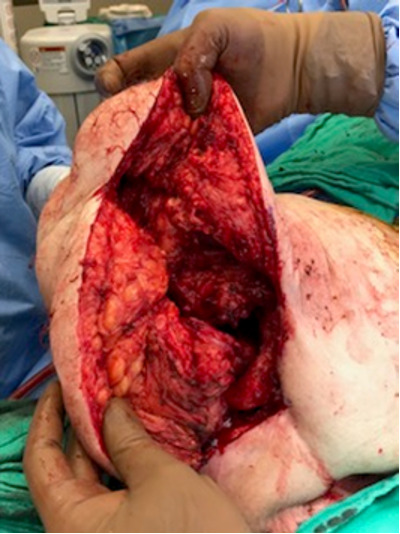
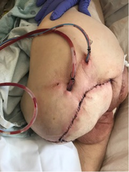
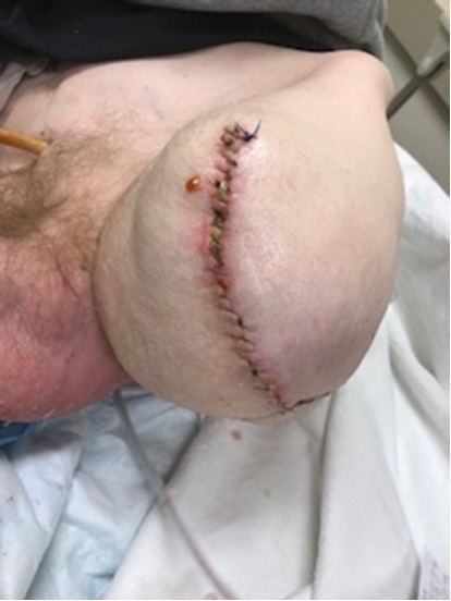
FIGURE 4.


a) Intraoperative view of the patient in a right lateral decubitus position. Previous left AKA site with osteomyelitis of left femoral head due to septic joint. b) Intraoperative view of the left HD prior to closure. c) The acetabular fossa was obliterated with vastus lateralis muscle flap and skin flaps closure achieved over 2 drains. d) Healing and intact incision at 2-week postoperative visit.
