Fig. 2.
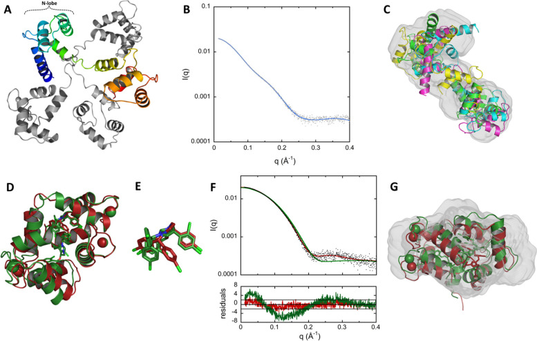
Structural models of holo-CaM using EOM and structures of holo-CaM:CDZ determined by X-ray crystallography. A Final ensemble of SAXS-derived conformations of holo-CaM using EOM. The four structural models (SASBDB ID: SASDNX3) are superimposed by aligning their N-lobe alpha carbons (residues 6 to 66) using Pymol. One structural model is shown in rainbow, the three others are in grey. B Comparison of experimental data (grey dots) to the calculated scattering pattern (blue curve) of the final EOM ensemble. C Fitting of the four EOM structural models of holo-CaM to the SAXS-derived DENSS volume. D X-ray structures of holo-CaM in complex with one (red, PDB ID: 7PSZ) and two (green, PDB ID: 7PU9) CDZ molecules. E Superimposition of the CDZ molecules of both structures showing the rotation of the chlorophenyl moieties. F Top: Fitting of the calculated scattering patterns of the two crystallographic structures of holo-CaM:CDZ obtained using Crysol to the experimental SAXS pattern recorded with 333 μM of CaM and 1050 μM of CDZ. The χ2 are 1.4 and 9.3 for the 1:1 (red) and 1:2 (green) holo-CaM:CDZ complexes, respectively. Bottom: Distribution of reduced residuals corresponding to the two fits presented above. G Fitting of the two crystallographic structures of holo-CaM:CDZ to the SAXS-derived DENSS volume (SASBDB ID: SASDNY3)

