Figure 1.
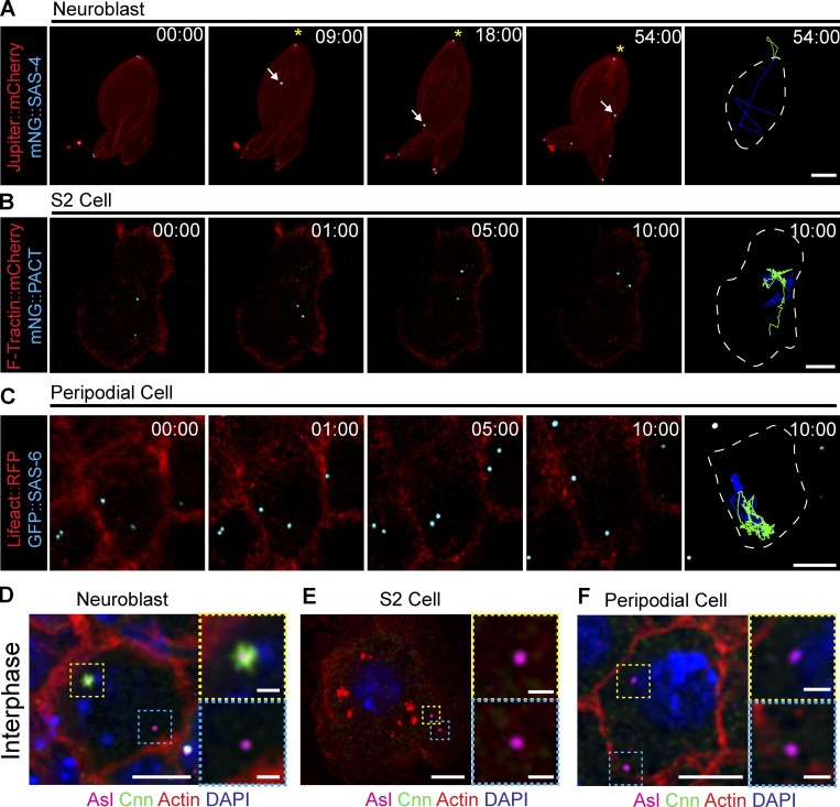
Drosophila centrioles are motile in interphase cells. (A) Z-stack projection of an interphase NB expressing Jupiter::mCherry (red) and mNG::SAS-4 (cyan). The mother centriole (asterisk) remains closely associated with the apical cell cortex; the daughter centriole (arrow) moves throughout the cell. (B) Cultured S2 cell transfected with F-tractin::mCherry (red) to visualize the cell and mNG::PACT (cyan) to label centrioles. Both centrioles are highly motile through the acquisition. (C) Peripodial cell expressing Lifeact::RFP (red) to visualize the cell and GFP::SAS-6 (cyan) labelling the centrioles. Both centrioles are highly motile within the cell. Last columns in A, B, and C show cell outline (white line) and 10-min time projections of centriole movement (green and blue lines). (D) Fixed NB showing that Cnn (green) is restricted to one of the two centrioles (magenta). (E and F) Fixed S2 cell (E) and peripodial cell (F) showing no Cnn (green) present on the centrioles (magenta). Scale bars: 5 µm; inset scale bars: 1 µm. Time stamp: mm:ss.

