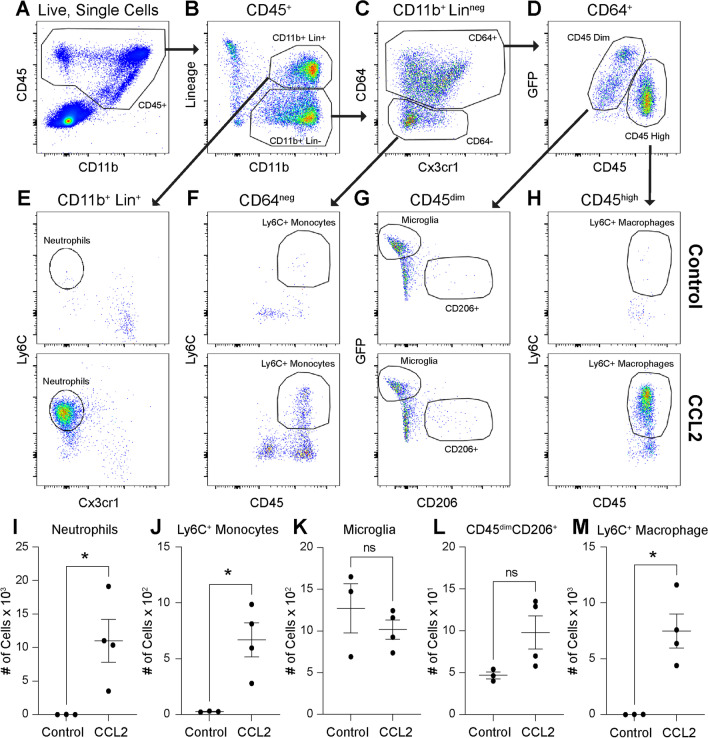
Fig. 7.
CCL2 injections increase inflammatory cells in the retina. The gating strategy for whole retina flow cytometry. All leukocytes are identified from live, single cells as CD45+ (A). CD45 + cells are stained with CD11b and Lineage (Lin) gate including CD4 and CD8 (T-cells), B220 (B-cells), NK1.1 (NK cells), SiglecF (eosinophils), Ly6G (neutrophils) to define CD11b+Lin+ and CD11b+Linneg cells (B). CD11b+Linneg cells were gated forward to delineate CD64+ and CD64neg cells (C). From CD64+ cells, CD45dim and CD45high cells were discriminated (D). Representative plots of CD11b+Lin+ cells from control and CCL2 groups identified Ly6C+Cx3cr1neg neutrophils (E). Representative plots of CD64neg cells from control and CCL2 groups defined Ly6C+CD45+ classical monocytes (F). Representative plots of CD45dim cells from control and CCL2 groups delineated GFP+CD206neg microglia and GFPnegCD206+ resident macrophages (G). Representative plots of CD45high cells from control and CCL2 groups discriminated Ly6C+ macrophages (H). CCL2 treatment increased neutrophil (I), classical monocyte (J), and Ly6C+ macrophage infiltration into the retina (M) with no change in microglia or GFPnegCD206+ macrophages (K–L). N = 3–4 mice (both eyes) per group from 2 independent experiments that were performed on two separate days from different litters and combined, *p < 0.05, two-tailed unpaired t tests with Welch’s correction

