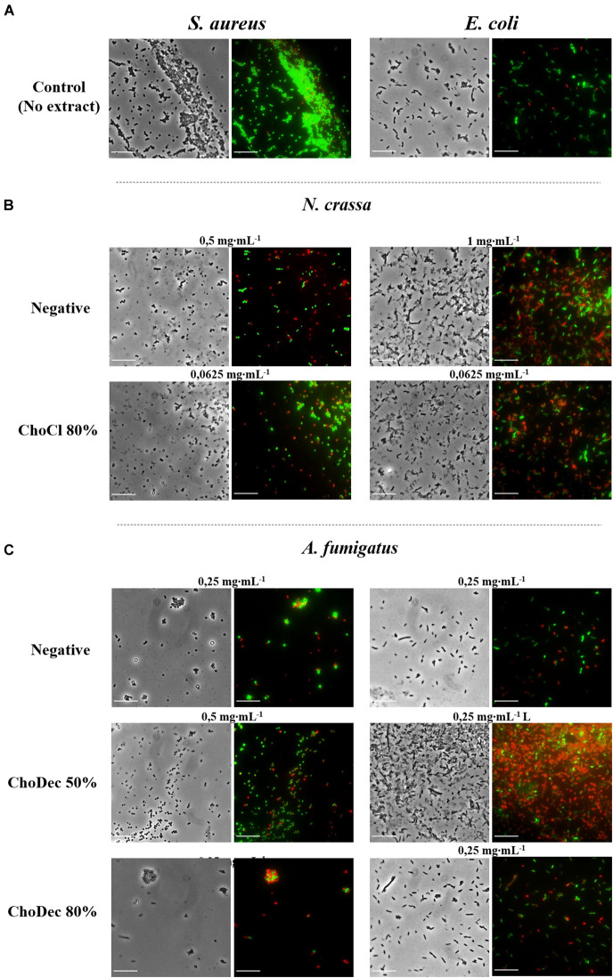
FIGURE 4.
N. crassa and A. fumigatus crude extracts led to significant lysis of S. aureus and E. coli cells, which is denoted by the red labeling. Microscopic snapshots of E. coli and S. aureus grown in the absence of extract (A) and in the presence of crude extracts derived from cultures grown in media with or without (i.e., negative) supplementation: N. crassa (B) and A. fumigatus (C). Images of bacteria at concentrations near the measured IC50 for each crude extract are shown. Cells were stained with SYTO9 (green) and PI (red) denoting live and dead cells, respectively. Scale bar, 10 μm.

