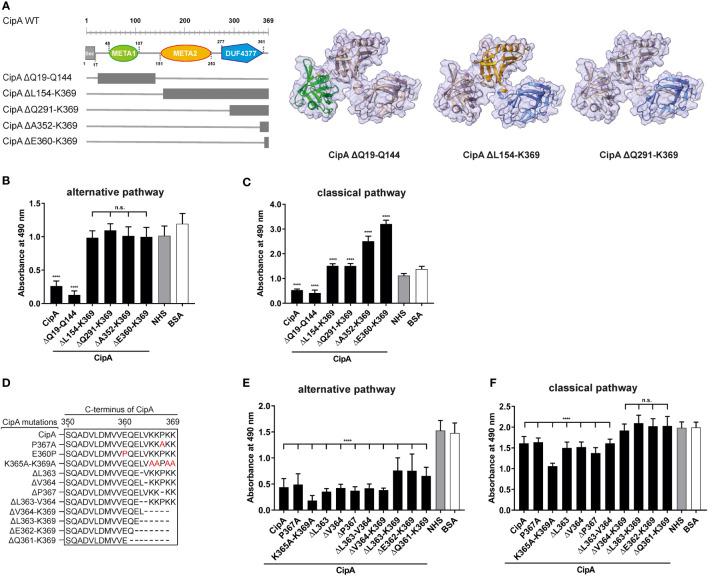
Figure 1.
Assessment of the inhibitory capacity of CipA and CipA variants on activation of the AP and CP. Schematic representation of N- and C-terminal deletion CipA variants and structure predictions generated by using AlphaFold2.0 (A). Deletions are as follows: CipA ΔQ19-Q144 removes the whole META1 domain (green), CipA ΔL154-K369 removes the META2 (orange) and DUF4377 (blue) domains, while the CipA ΔQ291-K369 deletion removes most of the DUF4377 domain. Schematic representation of CipA variants carrying single and multiple deletions or substitutions (in red letters) (D). WiELISA was performed to assess the inhibitory capacity of CipA deletion variants on the AP (B, E) and CP (C, F). NHS pre-incubated with the purified CipA proteins or BSA (500 nM for AP and 2.5 µM for the CP) were added to microtiter plates immobilized with LPS (AP) or IgM (CP). Formation of the MAC was detected by using a monoclonal anti-C5b-9 antibody. Data represent means and standard deviation of at least three different experiments, each conducted in triplicate. ****, p ≤ 0.0001, n.s., no statistical significance, one-way ANOVA with post-hoc Bonferroni multiple comparison test (confidence interval = 95%).

