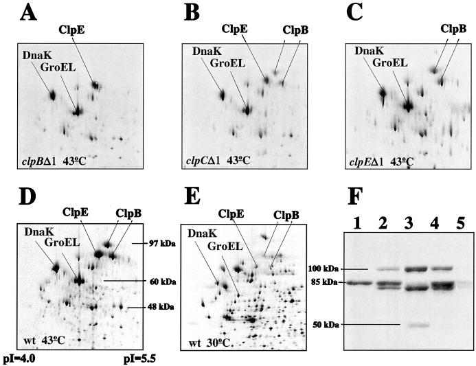
FIG. 4.
Western blot and two-dimensional protein analyses of Clp expression in wild-type (wt) and clp mutant cells. (A to E) 35S-labeled proteins extracted from equal amounts of cells of L. lactis MG1363 (D and E), HI1635 (clpBΔ1; A), HI1632 (clpCΔ1; panel B), and HI1615 (clpEΔ1; C) grown at 30°C (E) or shifted to 43°C for 20 min (A to D) were separated in two dimensions. (F) Proteins extracted from equal amounts of L. lactis MG1363 (lane 4 and 5), HI1635 (clpBΔ1; lane 1), HI1632 (clpCΔ1; lane 2), and HI1615 (clpEΔ1; lane 3) grown at 30°C (lane 5) or shifted to 43°C for 20 min (lanes 1 to 4) were separated on a sodium dodecyl sulfate–10% polyacrylamide gel and reacted with anti-Hsp104 antibody diluted 1:4,000. The molecular masses deduced from known proteins or protein molecular mass markers (Gibco BRL) are indicated, together with the pIs.

