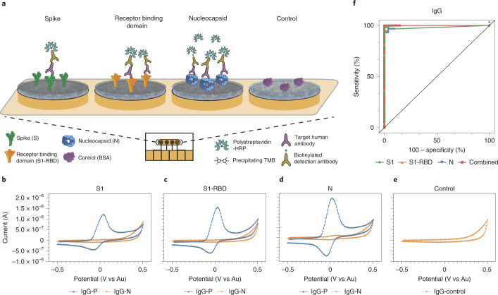
Fig. 3. Schematic and representative raw cyclic voltammetry data of the multiplexed serology assay.
a, Schematic illustrating the multiplexed EC serological assay to assess host antibody responses on electrodes functionalized with SARS-CoV-2 antigens. Host antibodies bind to the SARS-CoV-2 antigens immobilized on the chips. Subsequently, biotinylated anti-human IgG secondary antibodies bind, followed by polystreptavidin-HRP binding and TMB precipitation on the chips. b–e, Typical cyclic voltammograms for the four different electrodes that target host antibodies against S1 subunit (b), S1-RBD (c), N (d), and BSA negative control (e). f, ROC curves generated from the patient sample data obtained for the IgG EC serology assay.

