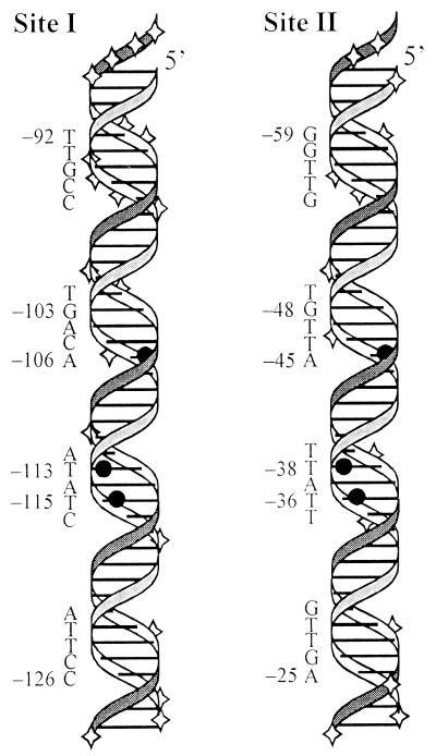
FIG. 6.
Three-dimensional representation of Rns binding sites I and II. The positions within each binding site that remain accessible or become hypersensitive to DNase I cleavage upon MBP::Rns binding are indicated by diamonds. Within each binding site the three thymine C5-methyl groups that MBP::Rns has hydrophobic interactions with are shown by solid circles. To align these thymines so that they appear in the same orientation in the figure, the sequence of binding site I has been inverted. The numbering is relative to the transcription start site of Pcoo.

