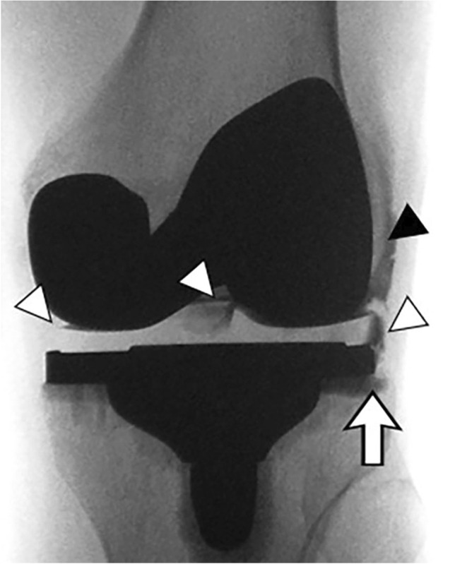
Fig. 14.

A 65-year-old female with painful total knee arthroplasty and suspected loosening. Fluoroscopic image after intraarticular contrast injection shows contrast outlining the polyethylene liner of the tibial tray (white arrowheads) and the lateral femoral condyle (black arrowhead). Contrast also extends into the metal-bone interface of the tibial tray (arrow) suggesting loosening of the prosthesis
