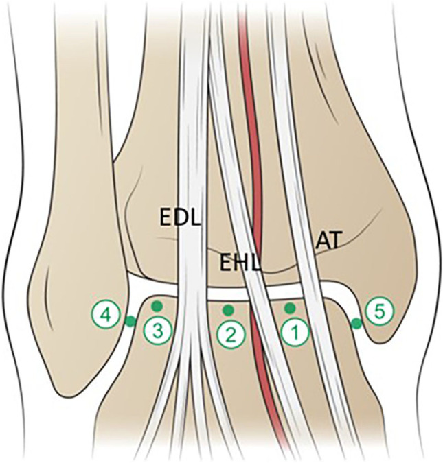
Fig. 16.

Illustration of ankle injection targets in the AP projection. 1 = Target is the medial talar dome between the AT and EHL tendons. 2 = Target is the central talar dome between the dorsalis pedis artery and the EDL tendons. 3 = Target is the lateral talar dome lateral to the EDL. 4 = Target is the upper half of the lateral clear space. 5 = Target is the upper half of the medial clear space. AT = anterior tibial tendon; EHL = extensor hallucis longus tendon; EDL = extensor digitorum longus tendons. Anterior tibial/dorsalis pedis artery is depicted in red
