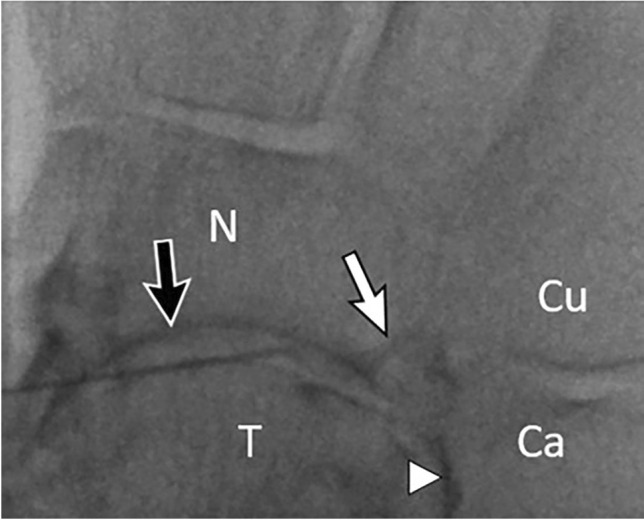
Fig. 25.

A 70-year-old female with history of tibiotalar fusion and midfoot pain, referred for talonavicular CSI. Fluoroscopic image shows contrast outlining the talonavicular joint (black arrow) and talocalcaneal joint (arrowhead). Contrast also outlines a calcaneal osteophyte at the calcaneonavicular articulation (white arrow). T = talus. N = navicular. Ca = calcaneus. Cu = cuboid
